


Breast cancer is most treatable when it is found early, and technology is steadily improving the odds of catching it in time. From artificial intelligence that reads mammograms alongside radiologists to 3D imaging and wearable research tools, detection is becoming faster, more accurate and more personal. With October marking Breast Cancer Awareness Month, it is a good moment to look at what is actually changing in clinics — and what patients should know about it.
Why Early Detection Matters So Much
Survival rates for breast cancer have improved dramatically over recent decades, and earlier diagnosis is one of the biggest reasons. A tumour found while it is small and confined is far easier to treat than one discovered after it has spread. The World Health Organization’s overview of breast cancer stresses the same point: organised screening, prompt diagnosis and access to treatment are the three pillars that save lives.
The challenge is that screening is imperfect. Mammograms can miss cancers, particularly in dense breast tissue, and they can also flag harmless changes that lead to anxious weeks of follow-up tests. Technology is now being aimed squarely at both problems — fewer misses, and fewer false alarms.
AI as a Second Pair of Eyes
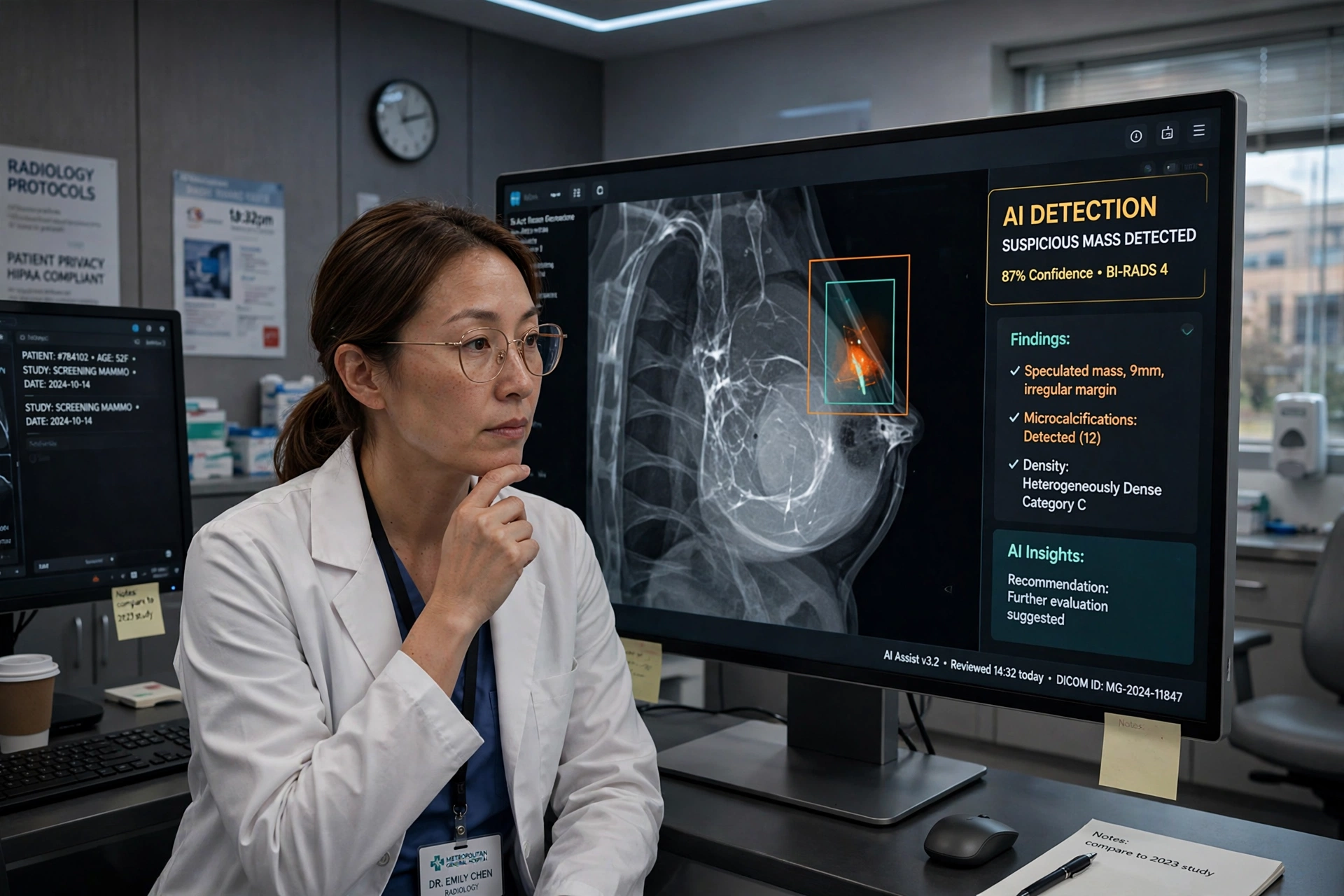
The most talked-about change is artificial intelligence in image reading. Modern AI systems are trained on millions of mammograms and can highlight subtle patterns that are easy for a tired human eye to overlook. In practice, most clinics use AI as a second reader or a triage tool: it flags the cases that need the closest attention and helps radiologists prioritise their workload.
Large studies in Europe and elsewhere have found that AI-supported screening can detect more cancers while reducing the number of women recalled for unnecessary follow-up. It is a similar pattern to the one we described in how on-device AI is quietly reshaping everyday technology — the software does not replace the professional, it makes the professional’s judgement faster and better supported.
3D Mammography and Better Imaging
Alongside smarter software, the imaging itself is improving. Digital breast tomosynthesis, often called 3D mammography, takes multiple images from different angles and reconstructs them into a layered view. Radiologists can effectively look through tissue rather than at a single flattened image, which helps separate overlapping structures that can hide — or imitate — a tumour.
Other tools are moving into wider use too. Contrast-enhanced mammography can highlight areas of unusual blood flow, ultrasound remains valuable for women with dense breasts, and MRI is used for those at higher risk. The National Cancer Institute maintains detailed, plain-language guidance on screening options and who benefits most from each, which is well worth reading before your next appointment.
Risk Prediction Is Getting Personal
Detection is only half the story. Newer AI models do not just look for cancer that is already there; they estimate a woman’s risk of developing it in the coming years, using patterns in images that humans cannot see. A higher risk score can mean earlier or more frequent screening, while a lower score may eventually mean safely spacing appointments further apart.
Genetic testing, family history tools and lifestyle data are increasingly combined into a single risk picture. That personalisation matters for everyday health decisions too — the same principle behind practical, family-focused care that we covered in the benefits of family dentistry for parents: know your risk, plan your check-ups, and do not wait for symptoms.
What Technology Cannot Do
For all the progress, no algorithm replaces regular screening or a prompt response to changes. AI systems can inherit biases from their training data, performance varies between populations, and access to the newest tools is uneven between — and within — countries. Regulators are therefore insisting on real-world evidence and human oversight, not just impressive laboratory results.
There is also a human side that machines cannot cover. A screening result, good or bad, arrives in a person’s life, not a dataset. Clear communication, fast follow-up appointments and support while waiting for results remain as important as any scanner.
What Patients Should Do Now
The practical advice is unchanged, but it bears repeating. Attend screening when you are invited, and know what is normal for you so you can report changes — a new lump, skin changes, or discharge — without waiting for the next routine appointment. If you have dense breasts or a strong family history, ask whether additional imaging or a personalised screening plan makes sense for you.
Technology is making breast cancer detection earlier, smarter and more tailored than ever before. The tools work best, though, when people actually use them. This October, the most powerful step is still the simplest one: book the screening, keep the appointment, and encourage the people you love to do the same.
